CASE 21: Complex primary hip replacement to reverse an arthrodesis in a mid-life woman
The Story
“I first met Helen at the age of 47 when she was referred to my clinic. She presented with worsening back pain and reduced mobility. As a young woman, Helen had suffered trauma to her right hip, which led to the joint being pinned and then fused with an arthrodesis plate.
The arthrodesis was done at the age of 18 years old: hip replacement would not have lasted long enough for her life. After extensive investigation of her back pain by the spinal surgeons, we deduced that her ongoing back pain was secondary to her arthrodesis, and was not related to any new spinal pathology.
She had coped with the stiff hip for many years but found the stiffness was becoming harder to cope with.”
The Investigation
On examination in clinic, Helen had an arthrodesed right hip with multiple surgical scars surrounding the joint. Despite the arthrodesis, the muscle bulk in the region of the hip abductors was clinically symmetrical to the left side. Her Oxford Hip Score for the right hip was 13/48.
The Evidence
Anteroposterior plain radiograph demonstrating fusion of the right hip joint with an arthrodesis plate. Her left hip joint looks healthy with minimal degenerative change.
Coronal MRI - We use the pre-operative MRI scan to evaluate the soft tissue and muscle bulk around the right hip. For Helen, this MRI demonstrates global muscle atrophy surrounding the right hip, which is to be expected with a hip fusion. Despite the atrophy, the abductors are present and with extensive physiotherapy would allow for a close to normal functioning arthroplasty.
Coronal CT - This scan was performed before the operation in order to aid in the planning of Helen’s surgery.
The Diagnosis
Helen was experiencing back pain and mobility issues because of her right sided arthrodesis fusing the joint.
The Plan
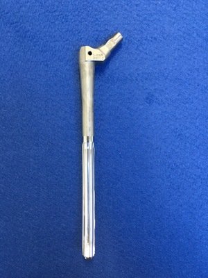
We made a plan to reverse Helen’s arthrodesis offering her conversion to a hip replacement. Helen was a very good candidate for an SROM femoral stem. This modular femoral component is an off the shelf system. It allows for the surgeon to select different components of the stem intraoperatively to allow for up to 10,398 different combinations to perfectly match the patients anatomy. Parameters such as the sleeve, stem length, proximal body type and head can be altered.
Trial components of the SROM
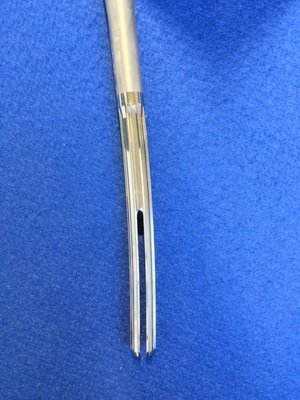
Final implant demonstrating a fluted distal segment for increased stability
Close up of the distal stem
The Operation
Helen underwent her arthrodesis reversal using a posterior approach through the old incision. The old plate was removed and an osteotomy performed.
The acetabulum was reamed to 49mm for a 50mm press fit socket, secured with two screws.
The femur was then prepared to accept an SROM stem, as seen in the images above. The joint was tested and stability achieved with a dual mobility bearing.
Gluteus medius was repaired. Although it was significantly wasted due to years of little use, it was still intact with the greater trochanter. The operation was finished with extensive washing and closure of the surgical site.
The Outcome
Anteroposterior plain radiograph showing that the SROM is in a satisfactory position the day after her operation.
Anteroposterior plain radiograph taken at Helen’s one year follow-up appointment. The implant hasn't moved from the immediate post-operative radiograph and there is evidence of bony healing.
Helen returned to our clinic one year after her procedure and with a lot of extensive physiotherapy to improve her muscle bulk, her mobility had improved massively. She was independently mobilising without any aids and her back pain had resolved.
On clinical examination she had a very good range of motion and she was Trendelenburg test negative. We were both very happy with the final result.
Trendelenburg test performed when Helen returned to clinic one year after her operation. Her pelvis is level meaning the test is negative. This is a good clinical indicator that the muscle bulk around her right hip has vastly improved.
The Verdict
“Reversal of a hip arthrodesis is a challenging clinical situation. Pre-operative work up, including the understanding of the patient’s expectation is important. In cases where the hip had formed normally (prior to their hip disease that resulted in arthrodesis) then there will always be an identifiable hip joint at surgery (soft tissue in the floor and familiar bony contours on the medial wall of the acetabulum).
CT helps plan the type, size and options of implants prior to surgery.
MRI helps with understanding the likely final result and stability: in the particular, the status of the abductor muscles predict the ability to walk, run and perform a negative Trendelenburg test.”
-
Functional impairment, and pain and degeneration of the neighbouring joints are frequent problems associated with a long-term fused hip: 50% of patients with fused hips complain of knee pain and low back pain. Indications to convert a fused hip to hip replacement include:
Disability resulting from a fused hip
Pain in the surrounding joints
Malpositioned arthrodesis
Painful pseudarthrosis
The procedure is technically demanding.
Complete or near complete relief is noted in 80% of patients with preoperative back pain and two thirds of patients with knee pain. Regarding pain and function in the surgically treated hip, 85% of hips are pain-free or with minimal pain, 80% have “good-to-excellent” range of motion. Survival of the converted THA ranges from 74% to 96% at 10 years and 73% at 26 years.
Between 75% and 100% of patients are satisfied with the procedure, and they generally are pleased with the outcome even if factors such as range of mobility, muscle strength, LLD, persistence of limp, and need of assistive walking aids are less satisfactory in converted hips compared with hips that had conventional primary THA.
A patient’s satisfaction with the obtained outcome may be related more to their overall improvement in quality of life rather than just the hip.
-
-
Please see Case 24 to learn the limitations of using modular-neck femoral stems


